Granulomatous slack skin: More than one type of mimicry

By Warren R. Heymann, MD
November 18, 2020
Vol. 2, No. 46

If Sir William Osler was alive today, he would need to modify his oft-quoted comment to: “He who knows syphilis, sarcoidosis, and CTCL, knows medicine.” What do all three disorders have in common? Granulomatous inflammation.
In their excellent review, Hodak and Amitay-Laish summarize the variants of mycosis fungoides (MF) that can imitate other disorders, leading to diagnostic dilemmas and confusion. Alphabetically, these presentations include bullous, folliculotropic, figurate erythema-like, granulomatous, hyperpigmented, hypopigmented, ichthyosiform, interstitial, other (non-specific), Pagetoid reticulosis, palmaris et plantaris, pigmented purpura-like, pityriasis lichenoides chronica-like, poikilodermatous, psoriasiform, pustular, and verrucous.
For improving the odds of diagnosing atypical cases of MF, the authors offer sage advice:
Maintain a high index of suspicion;
Perform a meticulous full body skin examination;
Discontinue topical steroids at least two weeks prior to obtaining skin biopsies;
Perform multiple biopsies from a variety of lesions;
If feasible, in equivocal cases, utilize the newly introduced high-throughput sequencing of the T cell receptor (TCR);
If necessary, continue to follow the patient and repeat biopsies periodically. (1)


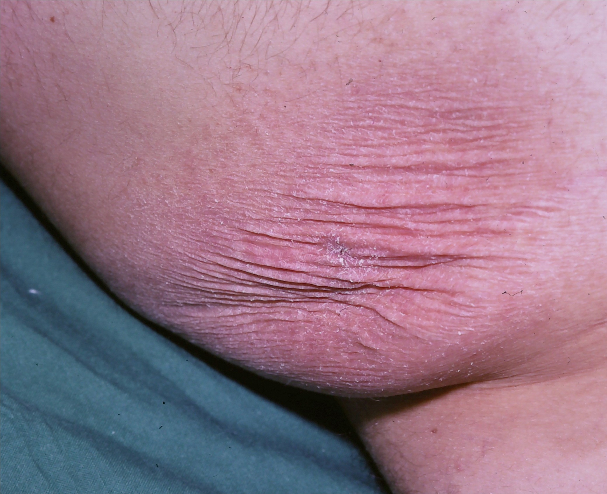
According to Wang et al: “Histopathologically, GSS presents with non-caseating granulomas with macrophagic and lymphocytic infiltrations. The distinction between GSS and granulomatous MF (GMF) can be challenging with merely pathological analysis. Elastophagocytosis occurs only in GSS and not GMF. However, it is rare finding in GSS and often missed by pathologists. Elastophagocytosis causes decreased or absence of elastic fibers in the dermis and results in clinically apparent skin laxity. Thus, elastophagocytosis is both a pathological feature and a key player in GSS pathogenesis.” The authors used electron microscopy (EM) to demonstrate that elastin fibers appear as amorphous masses. They observed fragmented elastin fibers in macrophages, indicating elastophagocytosis. These findings correlate with the disorganized elastin fibers characteristically observed with the Weigert elastic stain. (3)


If viewed in the broad context of granulomatous inflammation, the surprise is not that hypercalcemia is a complication, but why it is not observed more frequently. What protective mechanisms limit this complication in GSS and other granulomatous diseases to a minority of patients?
Point to Remember: Granulomatous slack skin is a rare variant of mycosis fungoides that may be complicated by hypercalcemia, as noted in other forms of granulomatous inflammation. Dermatologists should not be slack in systemic evaluation of patients with granulomatous disease.
Our Expert’s Viewpoint
Emmilia Hodak, MD
Chair, Division of Dermatology
Rabin Medical Center, Beilinson Hospital
Sackler Faculty of Medicine
Tel Aviv University
The David and Inez Myers Chair for Cancer Genetics
GSS is one of the many faces of mycosis fungoides (MF). Indeed, as Dr Heymann drew to our attention, all the three great imitators (syphilis, sarcoidosis, and MF) have in common granulomatous inflammation. However, in fact, granulomatous reaction is just one of all the other major histopathologic patterns encountered in inflammatory skin diseases (described by A. Bernard Ackerman, the greatest dermatopathologist of the 20th century), which can also be found in MF. The plethora of clinicopathological variants of MF is not unexpected, especially in the early stage; the number of malignant T cells is small and the dermal infiltrate consists mainly of reactive T lymphocytes, producing inflammatory cytokines that are partly responsible for the large repertoire of the histologic patterns observed in MF.
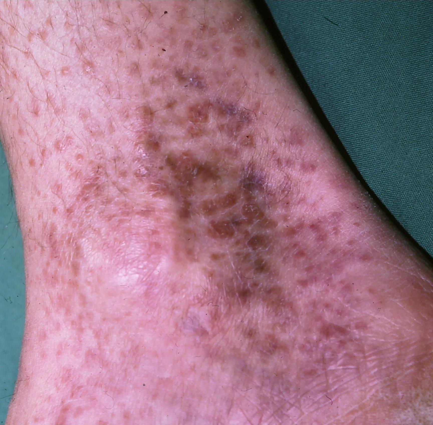
Various degrees of granulomatous reaction, as part of the inflammatory microenvironment, can be found in histopathological sections in about 4% of all cases of MF. This may be observed either at the time of the initial diagnosis or years later. Granulomatous MF is a rare variant defined by the European Organization for Research and Treatment of Cancer as MF which includes prominent granuloma formation, numerous histiocytic giant cells, or a histiocyte-rich infiltrate defined by histiocytes, accounting for more than 25% of the entire infiltrate. I have seen only two such patients in my entire career. Granulomatous MF is usually a histopathological variant detected in otherwise clinically conventional lesions of MF, and very rarely is a clinicopathological variant mimicking clinically benign granulomatous skin diseases, such as granuloma annulare, sarcoidosis, or granulomatous rosacea. GSS is a distinct clinicopathological subtype of granulomatous MF. In the very early stage, the clinical features may be misleading, as in the case presented above. However, in the course of disease the unique clinical features characteristic of GSS, with evolution to hanging skin folds, become apparent, and the clinical suspicion is quite straightforward.
The pathogenesis of granuloma formation in MF is unknown; however, it is recognized that macrophages are a major component of the infiltrate in the tumor microenvironment, playing a role in tumor development and progression in several cancers, including lymphoproliferative disorders. In MF it was found that the infiltration of macrophages typically increases during tumor progression from early to advanced stage. In line with these observations, there are some reports suggesting that patients with granulomatous MF experience more frequent disease progression than patients with classic MF. For some reason not yet investigated, this seems to be not the case in GSS, as the experience with such patients is that they usually have an indolent clinical course.
Hodak E, Amitay-Laish I. Mycosis fungoides: A great imitator. Clin Dermatol 2019; 37: 255-267.
Variants of mycosis fungoides. In Bolognia JL, Schaffer JV, Cerroni L, et al (eds). Dermatology, 4th edition, pp 2135.
Wang B, Zheng J, Wang HW. Granulomatous slack skin: Case report with electron microscopic features. Dermatol Online J 2019; 15: 25 (7).
Bettuzzi T. Ram-Wolff C, Hau E, de Masson A, et al. Severe hypercalcemia complicating granulomatous slack skin disease: An exceptional case. J Eur Acad Dermatol Venereol 2019; 33: e354-356.
Gwadera Ł, Białas AJ, Iwański MA, Górski P, Piotrowski WJ. Sarcoidosis and calcium homeostasis disturbances – Do we know where we stand? Chron Respir Dis 2019; 16:1479973119878713
Goltzman D. Nonparathyroid hypercalcemia. Front Horm Res 2019; 51: 77-90.
All content found on Dermatology World Insights and Inquiries, including: text, images, video, audio, or other formats, were created for informational purposes only. The content represents the opinions of the authors and should not be interpreted as the official AAD position on any topic addressed. It is not intended to be a substitute for professional medical advice, diagnosis, or treatment.
DW Insights and Inquiries archive
Explore hundreds of Dermatology World Insights and Inquiries articles by clinical area, specific condition, or medical journal source.
All content solely developed by the American Academy of Dermatology
The American Academy of Dermatology gratefully acknowledges the support from Incyte Dermatology.
 Make it easy for patients to find you.
 Meet the new AAD
 2022 AAD VMX
 AAD Learning Center
 Need coding help?
 Reduce burdens
 Clinical guidelines
 Why use AAD measures?
 Latest news
 New insights
 Combat burnout
 Joining or selling a practice?
 Advocacy priorities
 Promote the specialty

