Skin cancer types: Dermatofibrosarcoma protuberans signs & symptoms
Early signs and symptoms of dermatofibrosarcoma protuberans
This skin cancer tends to grow slowly, so it often goes unnoticed for months—or even years. When dermatofibrosarcoma protuberans (DFSP) first appears on the skin, a person may notice:
A pimple-like growth or rough patch of skin
No pain or tenderness where the growth or patch forms
Little change in the growth or patch
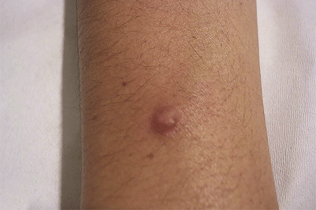
Dermatofibroma
DFSP often looks like this harmless and common skin growth, a dermatofibroma.

Dermatofibrosarcoma protuberans (DFSP) on a child’s skin
In children, this skin cancer tends to resemble a birthmark.

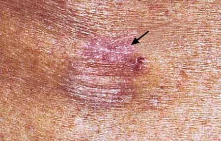
First signs of dermatofibrosarcoma protuberans (DFSP)
The first sign of this skin cancer may be reddish brown or pink patch of raised skin that looks like a scar.

As the skin cancer grows
As DFSP grows inside the middle layer of skin, it tends to push on the top layer of skin. You may see a lump, also known as a protuberan. The lump may feel hard or rubbery. As the lump grows, it stretches the skin. You may notice that the affected skin:
Becomes tender
Cracks and bleeds
Feels hard, and the lump seems cemented in the skin
When a woman is pregnant, DFSP tends to grow more quickly.
Over time, more protuberans (lumps) can appear. Once these appear, DFSP tends to grow quickly. In adults, the protuberans often range in color from reddish brown to violet. In young patients, DFSP tends to be blue or red in color.
As the dermatofibrosarcoma protuberans (DFSP) grows
Given time to grow, many protuberans can appear on the surface of the skin.

Where dermatofibrosarcoma protuberans forms on the body
DFSP can form anywhere on the skin. It is, however, more likely to develop on the:
Trunk (chest, back, abdomen, shoulder, buttocks)
Arm or leg
Few DFSPs form above the neck, but it is possible to find this skin cancer on the scalp or inside the mouth.
When to see a dermatologist
If you are worried about a growth on your skin, you should see a dermatologist. Many skin growths look alike. DFSP often looks like a harmless skin growth known as a dermatofibroma (shown above). This harmless skin growth rarely needs treatment. DFSP always requires treatment.
Dermatologists receive specialized training in diagnosing and treating skin cancer. This expertise is helpful when a person has a rare skin cancer like DFSP.
More common in younger people
DFSP tends to occur between the ages of 20 and 50.
Images
J Am Acad Dermatol 2010;62:247-56.
J Am Acad Dermatol 2009;61:1014-23.
J Am Acad Dermatol 2003;49:761-4.
J Am Acad Dermatol 2002;46:408-13
References
Buck DW, Kim JY, Alam M, “Multidisciplinary approach to the management of dermatofibrosarcoma protuberans. J Am Acad Dermatol. 2012;67(5):861-6.
Checketts SR, Hamilton TK, Baughman RD. “Congenital and childhood dermatofibrosarcoma protuberans: a case report and review of the literature.” J Am Acad Dermatol. 2000;42(5 Pt 2):907-13.
Criscione VD, Weinstock MA. “Descriptive epidemiology of dermatofibrosarcoma protuberans in the United States, 1973 to 2002.” J Am Acad Dermatol. 2007;56(6):968-73.
Gloster HM. “Dermatofibrosarcoma protuberans.” J Am Acad Dermatol. 1996;35(3 Pt 1):355-74.
Halpern M, Chen E, Ratner D. “Sarcomas.” In Nouri K. [editor]. Skin Cancer. United States. McGraw Hill Medical; 2008. p. 217-18.
Irarrazaval I, Redondo P. “Three-dimensional histology for dermatofibrosarcoma protuberans: case series and surgical technique.” J Am Acad Dermatol. 2012 Nov;67(5):991-6.
Kurlander DE, Martires KJ, Chen Y et al. “Risk of subsequent primary malignancies after dermatofibrosarcoma protuberans diagnosis: a national study.” J Am Acad Dermatol 2013;68(5):790-6.
Meehan SA, Napoli JA, Perry AE. “Dermatofibrosarcoma protuberans of the oral cavity.” J Am Acad Dermatol. 199;41(5 Pt 2):863-6.
Stivala A, Lombardo GA, Pompili G. “Dermatofibrosarcoma protuberans: Our experience of 59 cases.” Oncol Lett. 2012; 4(5): 1047–55.
Young RJ, Albertini JG. “Atrophic dermatofibrosarcoma protuberans: case report, review, and proposed molecular mechanisms.” J Am Acad Dermatol 2003;49:761-4.
 Think sun protection during Skin Cancer Awareness Month
 How to care for your skin if you have lupus
 Practice Safe Sun
 Sunscreen FAQs
 Fade dark spots
 Hidradenitis suppurativa
 Laser hair removal
 Scar treatment
 Botox
Botox
 Kids' camp - Camp Discovery
 Dermatologist-approved lesson plans, activities you can use
 Find a Dermatologist
 Why choose a board-certified dermatologist?